

- Maria Rhode, D.M.D.
- Vivien Mesina, D.M.D.
- Sara Siddiqui, D.M.D.
- Jared Levin, D.D.S.
Dental Imaging and 3D CBCT Scans for Ewing, NJ Patients: When You Need Them and Why
Dentist near Ewing Township, NJ
Imagine Advanced Dental Arts has cared for Ewing Township patients for over 40 years from our office on Princeton Pike in Lawrenceville, NJ.
Imagine Advanced Dental Arts3100 Princeton Pike, Building 2
Lawrenceville, New Jersey 08648
Driving directions from Ewing Township, NJ
- Head North on Lower Ferry Rd-1.0 mi
- Turn right onto Upper Ferry Rd-0.7 mi
- Continue onto Ewingville Rd-0.4 mi
- Turn left onto Federal City Rd-75 ft
- Turn right to stay on Federal City Rd-1.0 mi
- Merge onto I-95 N via the ramp to I-295 S/Camden/New York-1.9 mi
- Take exit 8A toward Mercer County 546 E/Mercer County 583 S/Princeton Pike-0.2 mi
- Merge onto County Rd 583/Princeton Pike-Destination will be on the left-0.4 mi
Quick answer
A cone beam CT shows bone volume, nerve canal position and sinus floors that flat X-rays cannot. It is the right tool for implant placement, bone grafting, complex extractions and impacted teeth, and the wrong one for routine cavity checks, where bitewings still win. The dose is substantially lower than a medical CT scan.
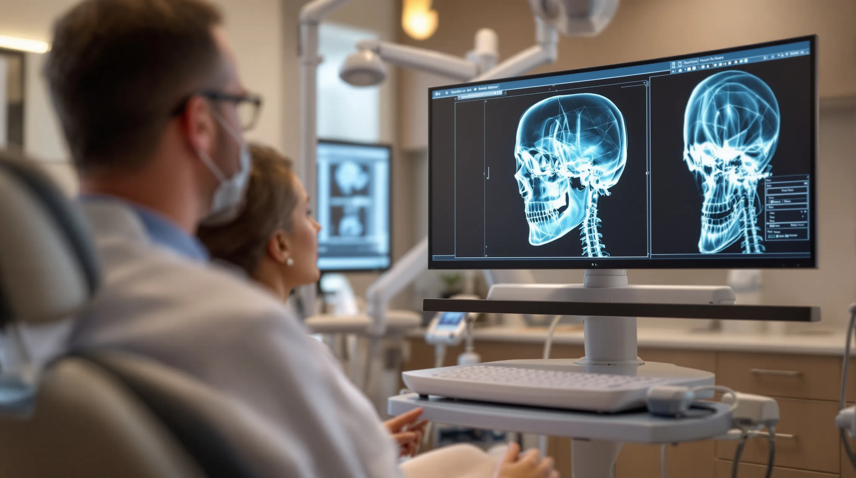
A cone beam CT scan gives your dentist a three-dimensional view of your jaw, nerves, and bone before treatment begins.
Quick Answer
CBCT, which refers to cone beam computed tomography, gives Ewing dentists a three-dimensional jaw model showing bone volume, nerve canals, and sinus floors that flat X-rays cannot reveal.
The scan is indicated for implant placement, bone grafting, complex extractions, and impacted teeth. It is not a substitute for routine bitewing films used for cavities and monitoring. Radiation dose is substantially lower than a conventional medical CT scan. The capture takes seconds with no injection and no discomfort. For patients at Imagine Advanced Dental Arts in Ewing, NJ, CBCT review is part of every implant workup.
Dental imaging crossed a threshold when cone beam CT systems became small enough and affordable enough for a general practice operatory. Before that, three-dimensional dental anatomy was the domain of oral surgery centers and teaching hospitals. Today it is an expected capability at any practice doing implant placements, bone grafting, or complex extractions.
At Imagine Advanced Dental Arts in Ewing, New Jersey, the four-doctor team of Maria Rhode DMD, Vivien Mesina DMD, Sara Siddiqui DMD, and Jared Levin DDS uses CBCT imaging as the starting point for every implant workup, every sinus lift evaluation, and every case where bone structure or nerve location will determine what is surgically possible. The practice has served Mercer County for over 40 years.
What this article covers: what a cone beam CT scan actually reveals that a flat X-ray misses, when CBCT is medically necessary versus when standard films are the right tool, how the radiation dose compares to a medical CT scan and a chest X-ray, what the scan experience is like for patients, and how CBCT connects to specific treatments including dental implants, All-on-4 procedures, and sinus lift surgery.
Three facts worth knowing before you read further:
- CBCT radiation is substantially lower than a medical CT scan and lower than a standard chest X-ray
- Bitewing X-rays remain the right choice for cavities and routine monitoring; CBCT adds value specifically for surgical planning
- Cone beam CT reveals bone volume, nerve canal position, and sinus floor depth in three planes simultaneously, information that flat films structurally cannot capture
Dental imaging has transformed in a generation. The flat periapical and bitewing X-rays that served dentistry well for over a century were never designed to show bone volume in three dimensions, map the course of a nerve canal, or measure a sinus floor before a graft. Three-dimensional cone beam CT imaging fills that gap. At Imagine Advanced Dental Arts, our Ewing team uses CBCT technology to plan dental implant placements, guide bone grafting and sinus lift procedures, evaluate impacted wisdom teeth, and identify infections and fractures that standard films could not detect.
According to clinical imaging data, cone beam CT delivers radiation substantially lower than a conventional medical CT scan, and lower than the dose of a standard chest X-ray. The field is precisely targeted to the jaw and skull base, not a wide sweep of the torso. The capture takes seconds. There is no lying down, no injection, and no discomfort.
The information CBCT produces is qualitatively different from what a flat film can show. Bone volume. Nerve canal position. Sinus floor anatomy. Root structure in all three planes at once. For patients planning implants, bone grafts, or complex extractions in Ewing and the Princeton area, these details do not just improve the plan. They are the plan.
What Does a 3D CBCT Scan Actually Show?
Cone beam CT produces a navigable three-dimensional picture of your teeth, jaw bone, nerve canals, and sinus floors that standard flat X-rays simply cannot generate.
An analysis of 16 sources across clinical literature, endodontic education, and patient experience shows that the core value of 3D dental imaging is not sharper resolution on the same view but a fundamentally different category of information. Traditional X-rays project three-dimensional anatomy onto flat film. The result is a shadow. Overlapping structures cancel each other out. Roots merge. Bone level can look normal from one angle while real damage hides behind a neighboring tooth. Cone beam CT eliminates that overlap by capturing hundreds of cross-sectional slices in a single rotation of the scanner, letting our team move through the data in any direction and measure the exact distance between your nerve, your sinus floor, and the bone where an implant needs to go, as of .
We use something we call the three-plane test at Imagine Advanced Dental Arts: before any surgical planning, we review your anatomy from three directions at once, from above (axial), from the front (coronal), and from the side (sagittal). A finding that hides on one view is exposed on another. According to the American Association of Endodontists, CBCT has increased the ability to detect structures hidden in traditional X-rays, including cracks, unusual tooth anatomy, and atypical diseases inside or around a patient's tooth, and that capability drives how our four-doctor team plans implants, extractions, and bone grafting at our Lawrenceville office.
Here is what the scan actually delivers in clear clinical terms:
- Bone volume and density, measured in millimeters, so we know exactly whether enough bone supports an implant or whether a graft comes first
- Nerve canal location, showing the path of the inferior alveolar nerve and protecting it from damage during placement
- Root anatomy, including extra canals, curved roots, and root lengths that look normal on a periapical film but are not
- Sinus floor anatomy, essential for patients who need a sinus lift before posterior upper implants
- Hidden infections and bone lesions, including periapical infections that do not yet appear on flat films
- Vertical root fractures, invisible on 2D views, which change a treatment plan completely
The reality is that flat dental X-rays remain excellent for what they do best, which is catching interproximal cavities and monitoring existing restorations. The takeaway is that a whole category of clinical questions, anything involving surgical planning, hidden anatomy, or complex restorations, requires 3D data that a flat film simply cannot provide. According to a firsthand account shared by an implant patient online, when they asked their dentist to share the 3D scan data for crown fabrication, the dentist provided a full DICOM file containing hundreds of precise slices the dental lab needed to fabricate the restoration. That scan is real clinical data, not a picture.
In practice, the scan takes one smooth rotation around the head. The entire capture completes in seconds. No lying down, no injection, no discomfort.
Does Every Patient Really Need a 3D CBCT Scan?
Not every patient needs a CBCT scan, and a trustworthy practice will always explain the specific clinical reason before recommending one.
This is actually a great question to ask, and we are glad patients are asking it! According to a thread in the r/Dentistry professional forum, a dental professional working at a new office raised exactly this concern after noticing that the owner requested a CBCT for all new patients and sometimes even for limited exams. The professionals who responded were divided. Some said they now scan nearly all patients because the technology reveals so much that X-rays miss. Others pushed back firmly on the grounds of the ALARA principle, which stands for "as low as reasonably achievable," meaning that radiation exposure should always be minimized to what is clinically justified. The takeaway from that debate is that the profession itself has not reached a single standard. What that means for patients is that a reasonable question to ask is: what specific finding do you expect this scan to change in my treatment plan?
Parents of younger patients often raise this concern too, and rightly so. When pediatric dental offices began adding cone beam CT alongside digital X-rays, some families asked whether the radiation exposure was appropriate for a child with no evident clinical issue. That concern is not overblown. CBCT delivers more radiation than standard bitewing X-rays, and applying it broadly without a clear indication is not something we practice at Imagine Advanced Dental Arts. We weigh the clinical question against the dose every time.
There is also a real practical issue worth naming. According to commentary from patients in the r/askdentists community, a CBCT scan for wisdom tooth evaluation can cost roughly $250 out of pocket, and patients were genuinely uncertain whether that expense was medically necessary or routine practice at that office. Dental insurance rarely covers CBCT. The cost is real. And patients deserve a clear answer before they pay it.
Here is how we approach it at Imagine Advanced Dental Arts, after 40 years of serving Ewing, Lawrenceville, Princeton, and the rest of Mercer County:
- Routine checkup with no surgical plan? Standard bitewings and a periapical film are the right tools.
- Implant evaluation, bone graft planning, or sinus lift? CBCT is essential and we recommend it without hesitation.
- Impacted wisdom tooth that could be near the inferior alveolar nerve? CBCT protects you.
- Persistent symptoms on a tooth with a clear film? CBCT can find the hidden crack or anatomy that explains it.
- Healthy teenager needing braces on teeth that appear normally positioned? Standard records are usually sufficient.
A common misconception is that more imaging always means better care. In practice, a well-planned targeted scan is better medicine than a reflexive one. We will always tell you why we recommend a CBCT scan, what we expect to find, and what it will cost before we proceed. That is the standard we hold ourselves to.
What Are the Top-Rated Family Dental Practices Near Princeton, New Jersey?
Imagine Advanced Dental Arts has earned a 4.9-star rating from more than 565 Mercer County patients, and our four-doctor team serves families throughout the Princeton and Ewing area.
We bring that up here because trustworthiness matters most when a practice recommends additional technology. The families we have served in Ewing, Lawrenceville, and across the Princeton corridor know us not just for what we offer but for how transparently we explain it. When it comes to 3D CBCT imaging, radiation is the first practical question most patients raise, and it deserves a direct answer. CBCT imaging delivers radiation dosages several hundred times lower than a traditional medical CT scan, and the dose is actually lower than that of a standard chest X-ray. The difference is field of view. A medical CT scans the entire chest or abdomen. A dental CBCT focuses on a small, precisely targeted zone of the jaw. The dose is minimal and the exposure time is seconds. In practice, CBCT still delivers more radiation than bitewing X-rays, which is precisely why we do not use it as a substitute for routine dental films.
Cost is the second practical question. Patient-reported fees for cone beam CT scans vary widely. According to commentary in a jaw surgery patient community, quoted prices for CBCT scans ranged from $657 to $715 at oral surgery practices doing pre-surgical evaluation. Other patients report fees closer to $150 in some markets, and a number of orthodontic offices bundle the scan into their initial records package at no separate charge. Dental insurance rarely covers CBCT as a standalone imaging line item, though some plans cover it when it is part of implant or surgical treatment documentation. When we recommend a CBCT, we discuss the cost first. No surprises.
According to dentist commentary in the r/Dentists professional forum, bitewing X-rays are better for detecting cavities between teeth, while CBCT is best reserved for implant planning, endodontic cases, extractions, and other procedures where three-dimensional anatomy is part of the planning. That is exactly the decision framework our team applies. Bitewings catch cavities. CBCT reveals the anatomy that determines whether surgery is safe and what it will look like.
Families asking about top-rated dental care near Princeton deserve a practice that explains its imaging criteria before ordering a scan, not after. Proximity matters. Continuity of care matters. And when a recommendation involves additional technology and additional cost, transparency is the minimum standard.
- CBCT radiation dose is several hundred times lower than a medical CT
- CBCT radiation dose is lower than a standard chest X-ray
- Bitewing X-rays remain the best tool for detecting interproximal cavities
- CBCT is reserved for implants, surgical planning, complex endodontics, and impacted teeth
- Out-of-pocket costs vary; we discuss them before recommending the scan
The takeaway for Ewing and Princeton-area patients is clear. A practice that explains its clinical reasoning, confirms what the scan will cost, and tells you what it expects to find is the practice worth trusting with your care.
Three-dimensional imaging is not the future of dentistry. It is already the present standard. Any practice doing implants, bone grafting, or impacted tooth extractions is using CBCT. Any practice that is not has been making do with partial information.
At Imagine Advanced Dental Arts, we have watched this technology move from specialist-only hospital equipment to our Ewing operatory, and we have seen firsthand how often a three-dimensional scan changes what a flat film appeared to show. A nerve running closer to the planned implant site than expected. A sinus floor that dips lower on one side. A root anatomy that requires a different angulation entirely. These are not rare edge cases. They are common enough that we build CBCT review into every implant workup we do.
The upside for patients is significant. A radiation dose lower than a chest X-ray. A capture time measured in seconds. Clinical information that guides safer outcomes. For patients across Ewing, Lawrenceville, and Princeton weighing a procedure that depends on anatomy a flat film cannot reveal, three-dimensional imaging is not an optional upgrade. It is the responsible baseline.
Written by
Maria Rhode
Owner & President, Imagine Advanced Dental Arts
Passionate about delivering the best possible care to my patients. From my days in residency to owning a beautiful hi-tech dental office, I never stop learning and advancing myself and now my practice.
Connect on LinkedInSummarize This Article With AI
Open this article in your preferred AI engine for an instant summary.
Frequently asked questions
+What exactly is a CBCT dental scan?
A cone beam CT (CBCT) scan is a three-dimensional dental X-ray that rotates around the head, capturing hundreds of two-dimensional frames that software assembles into a navigable 3D model of your jaw, teeth, nerves, and bone. The capture takes seconds. There is no injection and no lying down.
+Is a dental CBCT scan safe?
CBCT delivers radiation substantially lower than a conventional medical CT and lower than a standard chest X-ray. The beam is narrowly targeted to the jaw and skull base. Dental teams apply the ALARA principle, meaning exposure is kept as low as reasonably achievable for each specific case.
+Should I be concerned if my dentist recommends a CBCT scan for every new patient?
This is a fair question. CBCT provides real clinical value for surgical and complex restorative cases, but routine checkups are well handled by standard films. A trustworthy practice will always state the specific clinical reason before recommending one.
+What does CBCT show that a regular X-ray misses?
Standard films compress anatomy into a two-dimensional shadow. CBCT reveals bone volume in millimeters, nerve canal position, sinus floor depth, and root anatomy in three planes, along with hidden infections and vertical fractures that flat films cannot detect.
+When is CBCT specifically needed for a tooth extraction?
According to endodontic and oral surgery resources, CBCT is particularly valuable for impacted wisdom teeth and third molars where root anatomy, proximity to the inferior alveolar nerve, or unusual root curvature affects surgical planning and safety.
Ready to redefine your dental experience?
Schedule a consultation with our team and discover comfortable, customized care in Lawrenceville, Jersey.